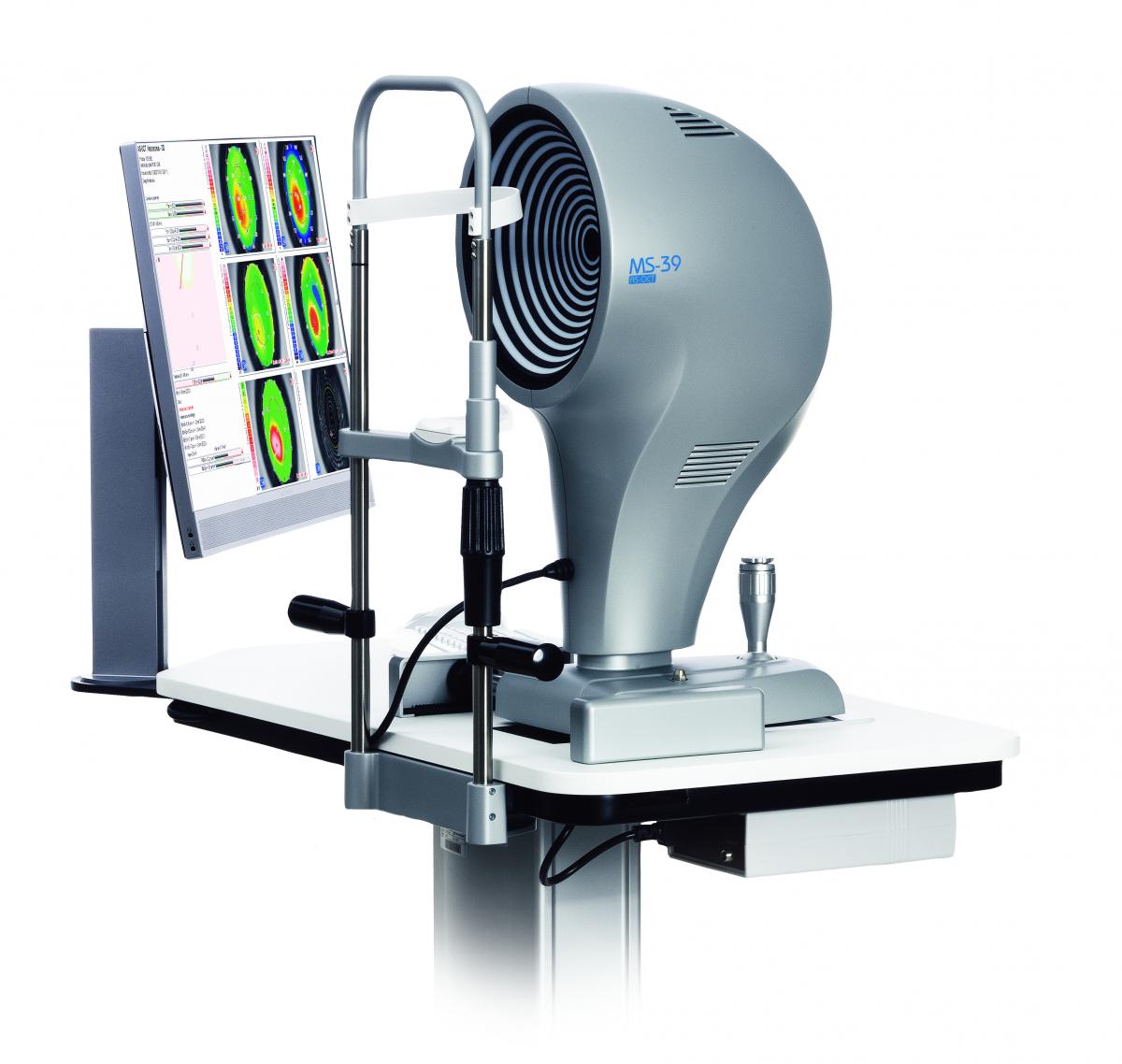
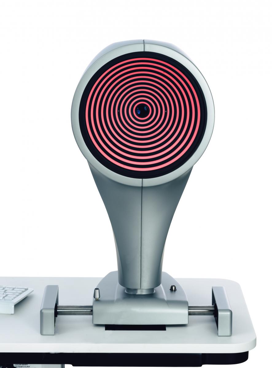
MS-39

AS-OCT

Is the most advanced device for the analysis of the anterior segment of the eye. MS-39 combines Placido disk corneal topography, with high resolution OCT-based anterior segment tomography. The clarity of the cross-sectional images, with a 16 mm diameter, along with the many details of the cornea structure and layers revealed by the MS-39, will be appreciated by  anterior segment specialists. MS-39 provides information on pachymetry, elevation, curvature and dioptric power of both corneal sufaces.

In addition to anterior segment clinical diagnostics, MS-39 can be used in corneal surgery for refractive surgery planning. An IOL calculation module is also available, based on Ray-Tracing techniques, Additional tools allow MS-39 to perform accurate pupil diameter measuremets and the advanced analysis of tear film.


CORNEAL SECTION

The sharpness of the high-resolution section images on a diameter of 16 mm, together with the many details of the structure and the cornea layers brought to light by the instrument, are the most extraordinary features and appreciated by the specialists of the anterior segment (corneal and epithelial) . The device provides pachymetry, elevation, curvature and power information for both corneal surfaces.

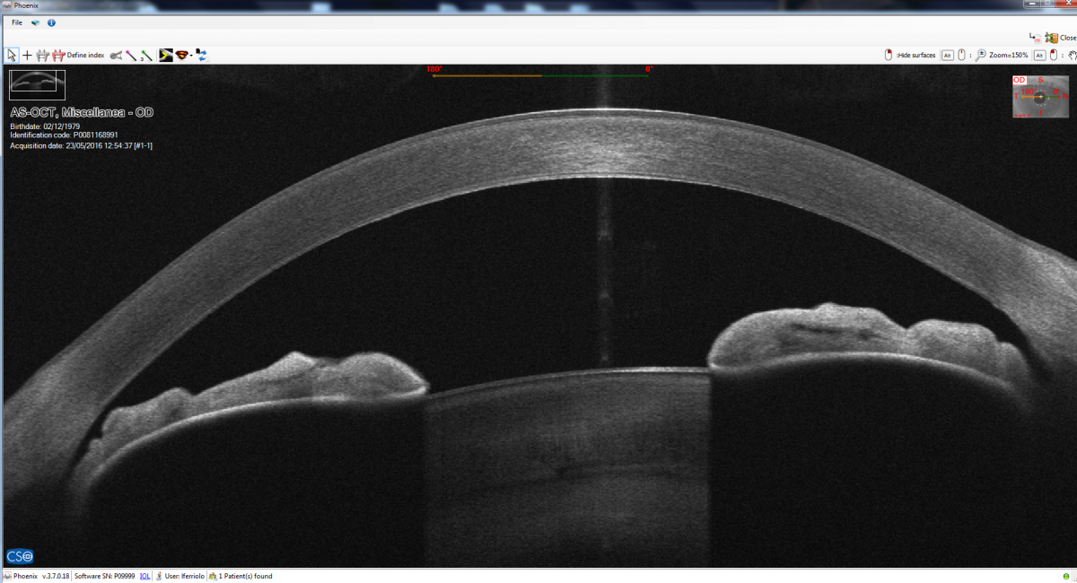
EPITHELIAL AND STROMAL MAP

MS-39 includes the advanced measurement of the epithelial and stromal layer. The epithelial masking effect is known, so knowledge of its morphology is very useful assess abnormalities of the corneal surface.  
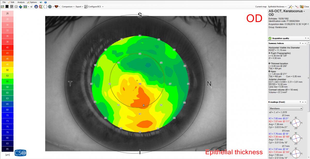
CORNEAL ABERROMETRY

Aberrometric analysis offers a complete overview of the corneal aberrations. It is possible to select the contribution of the anterior, posterior or total cornea for different pupil diameters. The OPD/WFE maps and the visual simulations (PSF, MTF, image convolution) can help the clinicain in understanding or explaining the patient’s visual problems.

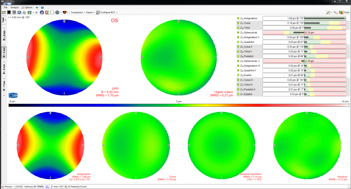
KERATOCONOUS SCREENING

Keratoconous screening provides the clinician with important information about the patinets cornea. Understanding this can help prevent complications associated with ectasia before corneal surgery is undertaken.  

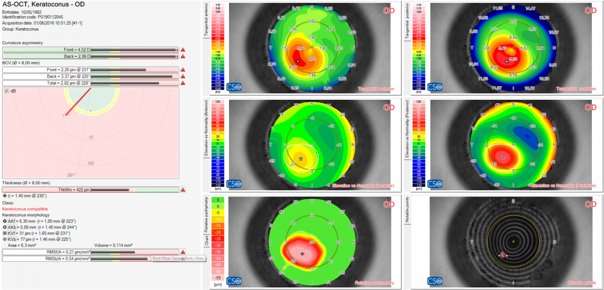
CRYSTALLINE BIOMETRY

In order to more accurately determine the ELED, and consequently to refine the intra-ocular lens calculation, MS-39 provides an acquisition mode to measure the crystalline lens thickness, its distance from the cornea and its equator.  
In vivo and in vitro results of an automated preloaded delivery system for IOL implantation in cataract surgery.
Mastropasqua, L, Toto, L, D'Ugo, E, Lanzini, M
Corneal graft rejection after yellow fever vaccine: a case report." Ocular Immunology
Author information
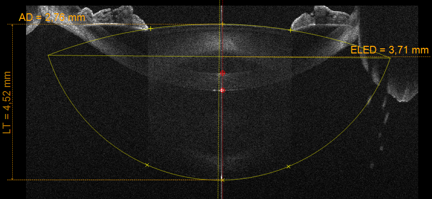
Vignapiano, R, Vicchio, L, Favuzza
Characteristics of endothelial corneal transplant rejection following immunisation with SARS-CoV-2 messenger RNA vaccine." British Journal of Ophthalmology, 2021
Author information:
Phylactou, M, Li, JPO and Larkin
Repeatability of automatic measurements by a new anterior segment optical coherence tomographer combined with Placido topography and agreement with …." Journal of Cataract & …, 2018
Author information
Savini, G, Schiano-Lomoriello, D and Hoffer, KJ.
Acute corneal endothelial graft rejection following COVID-19 vaccination." Journal Français d'Ophtalmologie, 2021, Elsevier
Author information
Crnej, A, Khoueir, Z, Cherfan, G and Saad, A.
Repeatability of anterior segment measurements by optical coherence tomography combined with Placido disk corneal topography in eyes with keratoconus
Schiano-Lomoriello, D, Bono, V, Abicca, I and Savini, G
Long-term endothelial cell loss with the iris-claw intraocular phakic lenses (Artisan®)
Author information
Galvis, V, Villamil, JF, Acuña, MF, Camacho, PA
In vivo microscopic and optical coherence tomography classification of neurotrophic keratopathy." Journal of Cellular
Author information
Mastropasqua, L, Nubile, M, Lanzini
Repeatability of automated measurements by a new anterior segment optical coherence tomographer and biometer and agreement with standard devices
Author information
Schiano-Lomoriello, D, Hoffer, KJ, Abicca, I and Savini, G
Corneal epithelial thickness intrasubject repeatability and its relation with visual limitation in keratoconus." American Journal of …, 2019
Author information
Vega-Estrada, A, Mimouni, M, Espla
Current developments in corneal topography and tomography
10. Kanclerz, P, Khoramnia, R and Wang, X
Agreement of white-to-white measurements with swept-source OCT, Scheimpflug and color LED devices.
Tañá-Rivero, P, Aguilar-Córcoles, S
Descemet membrane endothelial keratoplasty under failed penetrating keratoplasty without host descemetorhexis for the management of secondary graft failure.
Barrio, JLA Del, Montesel, A, Ho, V and Bhogal, M.
Corneal transplantation after failed grafts: options and outcomes.Corneal transplantation after failed grafts: options and outcomes.
Barrio, JLA Del, Bhogal, M, Ang, M, Ziaei, M
The use of anterior-segment optical-coherence tomography for the assessment of the iridocorneal angle and its alterations: update and current evidence.
15. Triolo, G, Barboni, P, Savini, G, Gaetano, F De
Epithelial and stromal remodelling following femtosecond laser–assisted stromal lenticule addition keratoplasty (SLAK) for keratoconus.
Nubile, M, Salgari, N, Mehta, JS, Calienno, R, Erroi, E
Femtosecond laser–assisted cataract surgery in eyes with shallow anterior chamber depth: comparison with conventional phacoemulsification.
Mencucci, R, Vitto, C De, Cennamo, M
Descemet membrane endothelial keratoplasty (DMEK) under previous DMEK for secondary endothelial graft failure.
Barrio, JLA Del, Vega-Estrada, A and Alió, JL.
Tear meniscus imaging by anterior segment-optical coherence tomography in medically controlled glaucoma.
Agnifili, L, Brescia, L, Scatena, B, Oddone, F
Graft detachment after Descemet's stripping automated endothelial keratoplasty in bullous keratopathy and Fuchs dystrophy.
Cardascia, N, Pastore, V, Bini, V
Machine Learning Algorithms to Detect Subclinical Keratoconus: Systematic Review.
Maile, H, Li, JPO, Gore, D, Leucci, M
Influence of age on small incision lenticule extraction outcomes.
Primavera, L, Canto-Cerdan, M, Alio, JL
Corneal cross-linking for keratoglobus using individualized fluence.
Hafezi, F, Torres-Netto, EA
Punctiform and polychromatic pre-Descemet corneal dystrophy: clinical evaluation and identification of the genetic basis.
Barrio, JLA Del, Chung, DD, Al-Shymali, O
Visual outcomes, footplate position and vault achieved with the Visian implantable Collamer lens for myopic astigmatism.
Reinstein, DZ, Vida, RS and Archer, TJ
Femtosecond laser-assisted deep lamellar descemet membrane endothelial keratoplasty for the treatment of endothelial dysfunction associated with posterior stromal
Barrio, JLA Del and Vargas, V.
Visual and Refractive Outcomes Following Laser Blended Vision With Non-linear Aspheric Micro-anisometropia (PRESBYOND) in Myopic and Hyperopic Patients.
Russo, A, Reinstein, DZ, Filini, O
National Risk Assessment of Impacts of Climate Change: Bridging the Gap to Adaptation Action
Stephen George Flood, Shona Paterson, Ellen O'connor,Barry O'Dwyer
Repeatability and comparison of corneal tomography in mild to severe keratoconus between the anterior segment OCT MS-39 and pentacam HR.
Seiler, TG, Mueller, M
The Precision of a New Anterior Segment Optical Coherence Tomographer and Its Comparison With a Swept-Source OCT-Based Optical Biometer in Patients With
Wang, Q, Chen, M, Ning, R, Savini, G
Efficacy and safety of Ab interno XEN gel implant after a failed filtering surgery.
Franco, FGS, Branchetti, M, Spagnuolo, V
Beauveria bassiana keratitis: Management of an atypical clinical presentation.
Ducange, P, Verdina, T, Stiro, F, Grottola, A
Diagnostics in Ocular Imaging
Mohammadpour, M and Heidari, Z
Epithelial remodelling masquerading as keratoconus progression: An interesting case report.
Gupta, K, Lalgudi, VG, Arora, V, Gupta, S
Demarcation Line Depth in Epithelium-Off Corneal Cross-Linking Performed at the Slit Lamp
Hafezi, F, Lu, NJ, Assaf, JF, Hafezi, NL, Koppen, C
Impact of Intraocular Lens Decentration on the Near Vision Performance of a Wavefront-Correcting Extended Depth of Focus IOL.
Nowrouzi, A, Barrio, JL Alió
A singular case of iatrogenic gas-filled double anterior chamber after DALK.
Franco, FGS, Vicchio, L, Branchetti, M
Applications of epithelial thickness mapping in corneal refractive surgery.
Reinstein, DZ, Archer, TJ and Vida, RS
Accuracy of the indices of MS-39 anterior segment optical coherence tomography in the diagnosis of keratoconic corneas.
Elkitkat, RS, Rifay, Y, Gharieb, HM
Phase retardation and corneal sublayer thickness repeatability using ultrahigh-resolution polarization-sensitive OCT
Patil, Rahul MTech; Shetty, Rohit MD, PhD; Patel, Yash MTech; Pisharody, Anchana MTech; Narasimhan, Raghav MTech; Pircher, Michael PhD; Hitzenberger, Christoph K. PhD; Nuijts, Rudy M.M.A. MD, PhD; Sinha Roy, Abhijit PhD
Combination of AS-OCT and Placido disc topography in keratoconus patient.
Bono, V
Repeatability assessment of anterior segment biometric measurements in myopic patients using a new anterior segment OCT with Placido corneal topography
Wang, H, Zhu, LS, Pang, CJ and Fan, Q
Uso clínico de MS-39: topógrafo para el estudio del segmento anterior.
Fortunato, Y Bauza, Salini, R Plasencia
Evolution of corneal epithelial remodeling after myopic laser in situ keratomileusis surgery measured by anterior segment optical coherence tomography
Díaz-Bernal, J, García-Basterra, I
Refractive Corneal Lenticule Extraction on Previous Photorefractive Keratectomy, with Optical Coherence Tomography Study.
Leccisotti, A, Fields, SV and Bartolo, G De
Epithelial Ingrowth Under LASIK Flap After a Massive Shockwave From the Port of Beirut Ammonium Nitrate Explosion.
Kheir, WJ, Assaf, JF and Awwad, ST
Multimodal ocular imaging in Proteus syndrome.
Salerni, A, Scartozzi, L, Piccinni, F
Customized Ablation Area PTK as a Treatment for Limewater-induced Calcium Carbonate Corneal Deposits.
Kozhaya, K, Chacra, LM
Anterior Segment OCT: Fundamentals and Technological Basis.
Vestri, G, Macaluso, C and Versaci, F.
Corneal epithelial thickness one year after myopic LASIK surgery, measured by anterior segment optical coherence tomography combined with Placido disk.
Díaz-Bernal, J, García-Basterra, I, Mora-Castilla, J
The corneo-scleral junction assessed with optical coherence tomography.
Muzyka-Woźniak, M, Oleszko, A, Stróżecki, Ł and Woźniak, S.
A Novel Microsurgical Robot: Preliminary Feasibility Test in Ophthalmic Field.
Savastano, A and Rizzo, S.
Phakic Intraocular Lens in Keratoconus.
Eldanasoury, A, Tolees, S and Bains, HS
IOL power calculation with ray tracing based on anterior segment OCT and adjusted axial length after myopic excimer laser surgery.
Savini, G, Hoffer, KJ, Ribeiro, FJ, Dias, JM
Clinical Outcomes Comparison of Combined Small Incision Lenticule Extraction with Collagen Cross-Linking Versus Small Incision Lenticule Extraction Only.
Chabib, A, Mammone, M, Fantozzi, C, Lian, RR
Amantadine therapy for Parkinson's Disease: In Vivo Confocal Microscopy corneal findings, case report and revision of literature.
Cennamo, M, Dragotto, F
Simultaneous Corneal Topography and Epithelial Thickness Mapping from a Single Measurement Using Optical Coherence Tomography.
Sikorski, BL.
Monolateral Epithelial Hyperplasia Caused by Boiling Oil Misdiagnosed as Unilateral Keratoconus.
Battaglia, B, Rosetta, P, Raimondi, R
Twelve-month outcomes of a new refractive lenticular extraction procedure.
Pradhan, KR and Mosquera, SA.
Descemet Membrane Endothelial Keratoplasty Under Failed Penetrating Keratoplasty Without Host Descemetorhexis for the Management of Secondary Graft
Ho, V and Bhogal, M.
3D Visualization System in Descemet Membrane Endothelial Keratoplasty (DMEK): A Six-Month Comparison with Conventional Microscope.
Morelli, A, Ferrandina, R, Favuzza, E
Delayed Laser In Situ Keratomileusis Interface Haze 6 Months After Corneal Cross-linking for Ectasia.
Assaf, JF, Hafezi, F and Awwad, ST
Corneal topographic changes after sequential suture removal in a corneal wound burn after phacoemulsification.
Pereira, S, Dorennavar, L, Nikhil, RP
Bilateral Small Incision Lenticule Extraction with SMILE pro using the VISUMAX 800.
Reinstein, DZ, CRS, PGD and Cert, LRS
Refractive Effect of Epithelial Remodelling in Myopia after Transepithelial Photorefractive Keratectomy
Ortueta, D de, Rüden, D von and Arba-Mosquera, S.
Vitamin D Supplementation Impacts Systemic Biomarkers of Collagen Degradation and Copper Metabolism in Patients With Keratoconus
Vitar, RML, Fonteyne, P, Knutsson, KA
Postoperative vault prediction for phakic implantable collamer lens surgery: the LASSO formulae.
Rocamora, L, Orlando, JI, Lwowski, C
Corneal ectasia part 3: OCT epithelial thickness measurements allow monitoring of corneal changes
Stewart, L.
A menetidôbecslés alkalmazásai.
Sándor, AM
SMILE for Hyperopia With and Without Astigmatism: Results of a Prospective Multicenter 12-Month Study.
Reinstein, DZ, Sekundo, W, Archer, TJ
Corneal Topography, Corneal Tomography, and Epithelial Maps in Keratoconus.
Reinstein, DZ, Archer, TJ, Vida, RS
National survey of corneal cross-linking (CXL) practice patterns in the United Kingdom during 2019.
Hayes, S, Jaycock, P, Rees, N, Figueiredo, FC
Linear Endotheliitis Following Posterior Chamber Phakic Intraocular Lens Implantation.
Barrio, JL Alió del and Alió, JL
Anterior Segment OCT: Clinical Applications.
Abdelghany, AA, Alió, JL, Barrio, JL Alió
Advanced epithelial mapping for refractive surgery.
Khamar, Pooja; Rao, Kavita1; Wadia, Kareeshma2; Dalal, Ritika3; Grover, Tushar4; Versaci, Francesco5; Gupta, Krati6
Diagnostic Value of Corneal Epithelial and Stromal Thickness Distribution Profiles in Forme Fruste Keratoconus and Subclinical Keratoconus. Cornea.
Toprak I, Vega A, Alió Del Barrio JL, Espla E, Cavas F, Alió JL.
Intraocular lens power calculation with ray tracing based on AS-OCT and adjusted axial length after myopic excimer laser surgery. J Cataract Refract Surg.
Savini G, Hoffer KJ, Ribeiro FJ, Dias JM, Coutinho CP, Barboni P, Schiano-Lomoriello D
IOL Power Calculation in Eyes Undergoing Combined Descemet Membrane Endothelial Keratoplasty and Cataract Surgery.
Knutsson, KA, Savini, G, Hoffer, KJ
Postoperative vault prediction for phakic implantable collamer lens surgery
Rocamora L, Orlando JI, Lwowski C, Kohnen T, Mertens E, Van Keer K.
Three-month outcomes of myopic astigmatism correction with small incision guided human cornea treatment.
Pradhan, KR and Arba-Mosquera, S.
Diagnostic value of corneal epithelial and stromal thickness distribution profiles in forme fruste keratoconus and subclinical keratoconus.
Toprak, I, Vega, A, Barrio, JLA Del, Espla, E, Cavas, F
Corneal stromal thickness changes after myopic laser corneal refractive surgery.
Barrio, JLA Del, Canto-Cerdan, M
Visian implantable collamer lens behavior in descemet membrane endothelial keratoplasty surgery.
Barrio, JLA Del, D'Oria, F and Alió, JL.
Corneal Epithelium Thickness and Refractive Changes After Myopic Laser Corneal Refractive Surgery.
Canto-Cerdan, M, Bahrawy, M El, Alió, JL
Coma influence on manifest astigmatism in coma-dominant irregular corneal optics.
Stojanovic, F, Reinstein, DZ
New Sizing Parameters and Model for Predicting Postoperative Vault for the Implantable Collamer Lens Posterior Chamber Phakic Intraocular Lens.
Reinstein, DZ, Archer, TJ, Vida, RS
Small Incision Lenticule Extraction (SMILE) for the Correction of High Myopia With Astigmatism.
Reinstein, DZ, Archer, TJ, Vida, RS
Intraoperative swept-source OCT–based corneal topography for measurement and analysis of stromal surface after epithelial removal.
Zhou, W, Reinstein, DZ, Archer, TJ
Epithelial Thickness Mapping for Keratoconus Screening by VHF Digital Ultrasound or Anterior Segment OCT.
Reinstein, DZ, Archer, TJ and Vida, RS

TECHNICAL DATA

Data transfer USB 3.0
Power supply external power source 24 VDC In: 100-240Vac - 50/60Hz - 2A - Out: 24Vdc - 100W
Power cable IEC C14 plug
Dimensions (HxWxD 505 x 315 x 251mm
Weight 10.4Kg
Chin rest movement 70mm ± 1mm
Minimum height of the chin cup from table 23cm
Base movement (xyz) 105 x 110 x 30mm
Working distance: 74mm
LIGHT SOURCES  
Placido disk illumination Led @635nm
OCT source SLed @845nm
Pupillographic illumination Led @950nm
TOPOGRAPHY  
Placido disk rings 22
Measured points 31232 (anterior surface) 25600 (posterior surface)
Topographic covering 10mm
Dioptric measurement range from 1D to 100D
Measurement accuracy Class A according to UNI EN ISO 19980-2012
TOMOGRAPHIC SECTION  
Image field 16mm x 8mm
Axial resolution >3.6µm (in tissue)
Transversal resolution 35µm (in air)
Image(s) resolution Keratoscopy (640x480) + 25 radial scans on a 16mm transversal field (1024 A-scan) - Section: on 16mm (1600 A-scan) on 8mm (800 A-scan)
MINIMUM SYSTEM REQUIREMENT  
PC: 4 GB RAM - Scheda Video 1 GB RAM (non condivisa) risoluzione 1024 x 768 pixels - USB 3.0 type A Sistema operati vo: Windows XP, Windows 7 e Windows 10 (32/64 bit).